Postgraduate Training Course in Reproductive Health/Chronic Disease
Ultrasonography: Recommendations for its appropriate use
in routine antenatal care in Nigeria
Dr Oladapo S. Sotiloye
Reproductive Health Care Centre
Department of Obstetrics and Gynaecology
Federal Medical Centre
Abeokuta, Nigeria
Supervisor: Margaret Usher-Patel
Implementing Best Practice Initiative
WHO/Department of Reproductive Health and Research
See also ![]() presentation
presentation
Abstract
Obstetric ultrasonography has grown in popularity since it was introduced over four decades ago. It had proliferated and its use if not controlled, which suggests that ad hoc use is open to misuse and could be a drain on the limited human and economic resources available without corresponding improvement in fetal and maternal outcomes.
The objective of this paper is to review current literature on the appropriate use of ultrasound during routine antenatal care and make recommendations for the appropriate use of ultrasound in Nigeria. The concern is that the Federal Republic of Nigeria is the largest black nation in the world with a vast population. Despite the amount of human and material resources available, the majority of the population are still living below the poverty line. Only 50% of the women receive antenatal care and if scarce resources are wasted on unnecessary equipment and the performance of scans by unskilled practitioners, technical, human and financial resources are wasted that could be used to increase access to the services and improve maternal and neonatal outcomes.
Extensive search mechanisms available through the WHO Headquarters electronic resources (The OVID, Medline, Popline and Cochrane data bases) were searched and other sites, such as the Blackwell SYNERGY and on-line journals. A manual search was also conducted, and Google search to determine the type of information available to the lay public. 54 papers were obtained for review.
Four systematic review were identified, three of them Cochrane reviews, eight randomized controlled trials and a number of other studies were reviewed. The main findings were that routine ultrasound has not resulted in improved fetal and maternal outcome when compared with the selective use (only when clinically indicated). The cost of routine ultrasound is high and the unnecessary human and material resources could be channelled to other useful areas.
The conclusions reached is that
ultrasound can be a useful tool in the antenatal period when employed judiciously based on sound clinical judgement. It should not be used indiscriminately. It should be used by trained, accredited and regulated practitioners. National guidelines should be available to guide use and inform both the practitioner and client. The recommendations made should support the further study of this issue in Nigeria and the formation of a national working group to develop national standards and protocols of practice.
Introduction
Obstetric ultrasonography has grown in popularity over the last thirty years and is fast becoming a standard component of antenatal care. In routine antenatal care it is used mainly to check the normal progression of pregnancy and identify deviations from the norm. In principle it is being used as a modern diagnostic and monitoring aid to undertake timely and appropriate clinical interventions to reduce maternal and perinatal morbidity and mortality. This paper questions whether the misapplication of this effective technology may however end up increasing the cost in all its ramifications (human technical and material resources) of routine antenatal care to all stake-holders without corresponding improvement in fatal or maternal outcome.
The diagnostic ultrasound employed in medicine is a sophisticated electronic technology which utilizes pulses of high frequency sound waves (>20,000 Hz) which is undetectable by the human ear (1,2). The transducer emits pulses of high frequency sound waves which pass through the tissue and some are reflected back at difference tissue interphase. The intensity of the reflected echo depends on the characteristics of tissue on the path of the propagated sound wave. Conductivity of ultrasound increases with the density of the material/tissue. Therefore, the velocity of ultrasound varies through various body organs with air having the slowest velocity of about 331 meter per second and bone having a much faster velocity of 4,080 meter per second (2).
The reflected wave is picked by the same transducer and converted into electronic signals for computer processing before it is displayed on the screen based on its intensity and position. The spatial relationship of the multiple displays forms the real-time images seen on the screen.
The technology of ultrasound has progressed from the earlier large and cumbersome models, which were modifications of military Sound Navigation and Ranging (SONAR) devices used
for submarine military operations to the more sophisticated real-time, 3-dimensional and the doppler flow sonography in use today (3). The earlier applications were in therapy rather than diagnosis utilising its heating and disruptive properties at higher doses (frequency and duration related) in physical and rehabilitation therapy, and neurosurgical ablations (4).
First application in diagnostic medicine was in neurosurgery in the early 1940s (5), later the unidirectional A-mode was used for tissue diagnosis of surgical materials (intestine, breast, etc) (5). Application to living tissue developed over the next ten years with what was termed a safe range of sound frequency (20,000 - 20,000,000 Hz). Today only 3-10 MHz are employed in clinical practice (6,7,8) . Ultrasonography was introduced into obstetrics through the pioneering work of Professor. Ian Donald in Glasgow, 1958 (9).
At that time, the ultrasound equipment was handled mainly by people involved with its development and their associates who were adequately exposed to its functionality and competent in its use.
In Nigeria, the technology was introduced about 30 years ago. For a long time, it was found in only one centre, University College Hospital, Ibadan and it was handled by highly skilled professionals. The advances in technology has made the ultrasound machine small and portable. Although it remains very expensive, nowadays it is possible to find this equipment in virtually every nook and cranny of the country.
Initially, it was in the purview of the Radiologists. As the popularity of ultrasound increases so is its use and now more and more obstetrician and general practitioners are using ultrasonography in routine antenatal care.
Many of these machines are operated by individuals who are either inadequately trained or have received no training at all. Up till the moment of this review ultrasonography has no definite place in the undergraduate curriculum. Attempts
are being made to entrench it into the postgraduate training, but this has still not been formalized or standardized. Those who were involved with obstetric ultrasonography either had a formal postgraduate training or were trained on the job. Majority of those who are actually practising ultrasonography now either had ad-hoc training or no training at all.
Training appears to be a none issue. A quick search of literature published over the last twenty years in Nigeria identified 61 papers written on the use of ultrasound (PubMed - National Library of Medicine). None of these papers addressed the issue of standardization or guidelines for standard practice. Most were focused on the fetal assessment and the diagnosis of anomalies.
The unsuspecting pregnant woman accepts the procedure without due consideration to the fact that the quality of ultrasound imaging does not only depend on the sophistication and resolutions of the equipment alone, but also on the experience and expertise of the operator (1,9 ,10,11) . Mistakes could occur at any level, but the frequency of mistake tends to reduce with expertise and experience (11,12) .
Moreover, from the literature reviewed it would appear that routine ultrasonography has not been found to result in improved fetal or maternal outcome (13,14) . The resources used on unnecessary ultrasound scans may be better utilised elsewhere in the health services (15) or the patient's home.
To use ultrasonography to provide our clients with the most appropriate and effective level of obstetric care, it is important that not only are practitioners trained in the appropriate use of this equipment, but also its use is governed by guidelines. Moreover, to protect the rights of the mother and unborn child, the level of performance should be periodically audited and governed by a regulatory body (15).
Objectives
- To review current literature to determine the appropriate use of ultrasound during routine antenatal care.
- To identify a series of recommendations that can be used to guide the appropriate use of ultrasound during routine antenatal care in Nigeria.
Methodology
Literature search for this review employed many different channels. The OVID database was searched through the WHO Headquarters electronic resources. The Medline, Popline, and the Cochrane databases were also searched using several approaches.
The Lancet was obtained on-line through the WHO Intranet and also the Headquarters electronic resources.
The Blackwell SYNERGY was also searched and some details obtained from on-line journals through the WHO Headquarters electronic resources subscriptions. The Googles search was used mainly to identify information available to the general public.
Manual search of relevant journals was also done.
The keywords and phrases used in the search include:
- Ultrasonography
- Ultrasound in pregnancy
- Routine ultrasound in pregnancy
- Ultrasound in early pregnancy
- Ultrasound in late pregnancy
- Cost and effectiveness AND obstetric ultrasound
- Routine ultrasound and fetal outcome
- Fetal outcome
- Maternal outcome
- Fetal anomaly scan
- Ultrasound in pregnancy AND safety.
Country Profile
Federal Republic of Nigeria is the largest black nation in the world and the population is projected to be about 120 million (projection from 1991 census) . It was however estimated to be 116,928,000 in 2001 by the UNFPA and WHO.
The budgetary allocation to health is far below the minimum recommended proportions of the national budget of 5-7%; it ranged between 1.5% to 2.5% of the national budget (16).
Per capital income is low, the gross domestic product (GDP) was $884.00 at the international rate but when the prevailing exchange rate in 2001 is taken into consideration it will come to $350.00 (or even lower 2 years later). The estimated government expenditure on health is about 2.2% of the GDP. The average standard of living is quite low.
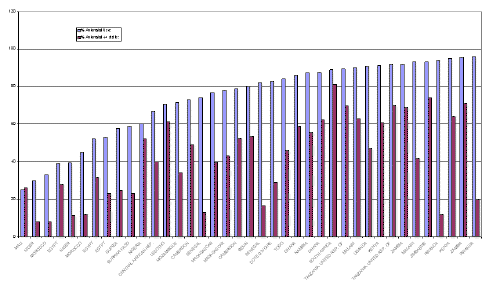
Despite this, the fertility rate is still high - 5.6% (17). Obstetric cost is not borne by the employer or government so any extra cost must be meaningful and worthwhile for the poor family. From figure 1 it is possible to determine that on average approximately 60% of Nigerian women receive ante natal care and 50% of these women will make at least 4 or more ante natal visits during their pregnancy (18). If ultrasound is used at every visit this has huge implications for the use of resources that might better be employed reaching the 40% of the population that receive no antenatal care. At the Federal Medical Centre, Abeokuta, the average number of ante-natal visits per woman is 6, but can range from 1 to 14. Ultrasound is used selectively when medically indicated.
Figure 1: Percentage of women with 4+ ante natal care visits in 20 African countries.
Results of the Review
From the literature reviewed it would appear is if there is no improvement in the outcome of pregnancy with the use of routine ultrasound (18). The weight of evidence is in favour of the use of ultrasonography only when clinically indicated (19-20). Routine ultrasonography can only be justified in areas where the incidence of fetal anomaly is high and pregnancy termination, at least before 24 weeks, is allowed legally (2) and morally.
The literature defines many valid uses for diagnostic ultrasonography and verifies the indications for use in pregnancy. There is a case for judicious use in pregnancy and in situations where the prevalence of pelvic infection is high with resultant pelvic adhesions. It is also useful for confirming early pregnancy and determining the site of implantation early enough to diagnose ectopic pregnancy before rupture (21).
The need for appropriate level of training, accreditation and regulation of the practice of ultrasonography is an area that needs further investigation so that the practice can be standardised and quality of care assured. The literature has been used to define a framework for recommending a standard of use in Nigeria for routine antenatal care.
Critical Analysis and Discussion
When to use Ultrasound in Early Pregnancy?
WHO has undertaken a randomised controlled trial of routine antenatal care and as a result recommends four visits during pregnancy (24). Three systematic reviews on the use of ultrasound in pregnancy discouraged routine use, but did not specify frequency of use or recommend when ultrasound should be used (1,10,22,23). These reviews focused on the clinical use of ultrasound, which in itself can help to define appropriate time frames for use.
As previously stated the usefulness of ultrasound in pregnancy, when clinically indicated, has rarely been questioned. The controversy has mainly been over the routine use of ultrasound in all pregnancies(3).
As noted by Neilson in the Cochrane Review on Ultrasound for Fetal Assessment in Early Pregnancy (2002) (10) there are a number of indications for its use in early pregnancy. These include more accurate calculation of gestational age, early identification of multiple pregnancy and diagnosis of non-viable pregnancies, early bleeding, inappropriately grown (growth restriction or macrosomia) and certain other fetal malformation.
Complications can however arise in pregnancy without clear warning signs or risk factors. For this reason there is a case for routine screening in pregnancy (early, late or both). Earlier diagnosis of ectopic pregnancy is also worthy of note here especially in combination with serial or critical beta hCG levels.
Using ultrasound to confirm pregnancy is not a difficult task, however, to make appropriate diagnosis of complications of early pregnancy can be challenging in inexperienced hands. A lot depends on the quality of the machine and the experience and expertise of the operator. Routine ultrasonography in the hands of inexperienced operator could do more harm than good (10). Findings of unclear significance may add to the anxiety and distress of the parents-to-be and lead to mismanagement (13).
Common applications of ultrasound in early pregnancy
Ultrasound provides the anatomic dimension of early pregnancy growth and confirmation of pregnancy (26). The cost of the procedure will however still be far more expensive than the standard simple urinary pregnancy (hCG) test following a missed period. The advantage of ultrasound in the confirmation of viability and localisations of the implantation site should possibly only be considered when a complication such as ectopic pregnancy is suspected.
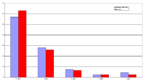
Early ultrasound gestational age dating is generally believed to be more accurate (25,26). A multi-centred study undertaken Campbell et al conducted on 4,527 women in 1985 found that there is no clear evidence that this, when done routinely, will lead to fewer overall inductions rate or contribute significantly to improved fetal outcome (31). Figure 2 provides a summary of their findings.
Figure 2. Percentage of patients delivering spontaneously within (+/-) one, two, three, four and > four weeks of estimated date of delivery based on the last menstrual period and ultrasound at 12 - 18 weeks.
Ewigman et al (1993) (18) conducted a randomised controlled involving 15,151 pregnant women at low risk for perinatal problems to determine if routine use of ultrasound decreased the frequency of adverse pregnancy outcomes. The conclusion reached was that routine use of ultrasonography did not improve perinatal outcomes as compared with selective use. A similar conclusion was reached even for multiple pregnancies. In this study, there were 68 and 61 multiple pregnancies in the ultrasound screening group and control group respectively. The rate of adverse outcome of 25% in the ultrasound screening group was not significantly different from 37.7% in the control group (Relative risk0.7, 95% CI, 0.39 to 1.11). Again, this study demonstrates that routine use of ultrasound does not improve outcome in multiple pregnancy despite early diagnosis. Table 1 shows the rate of adverse perinatal outcomes of 5.0% ultrasound group and 4.9% in the control group. (Relative risk 1.0, CI 0.9 - 1.2).
Table 1. Adverse Perinatal Outcomes in the Ultrasound-Screening and Control Groups
| Outcome | Singleton fetuses | Multiple gestation | All | |||
| Ultrasound screening | Control | Ultrasound screening | Control | Ultrasound screening | Control | |
| N = 7549 | N = 7473 | N = 136 | N= 123 | N =7685 | N=7596 | |
| Fetal death | 31 (0.4) | 22 (0.3) | 3 (2.2) | 1 (0.8) | 34 (0.4) | 23 (0.3) |
| Neonatal death | 17 (0.2) | 15 (0.2) | 1 (0.7) | 1 (2.4) | 18 (0.2) | 18 (0.2) |
| Severe morbidity | 88 (1.2) | 82 (1.1) | 11 (8.1) | 13 (10.6) | 99 (1.3) | 95 (1.3) |
| Moderate morbidity | 215 (2.8) | 213 (2.9) | 17 (12.5) | 24 (19.5)* | 232 (3.0) | 237 (3.1) |
| All adverse outcomes | ||||||
| Fetuses | 351 (4.6) | 332 (4.4) | 32 (23.5) | 41 (33.3)* | 383 (5.0) | 373 (4.9) |
| Pregnancies | 351 (4.6) | 332 (4.4) | 17 (25.0) | 23 (37.7) | 368 (4.8) | 355 (4.7) |
Vaginal bleeding during early pregnancy is one of the indications for ultrasonography (2,10,26,28). Viability can be determined and the different varieties of abortions diagnosed. Molar pregnancy is one of the differentials that could be diagnosed on sonography. Any associated pelvic mass could also be evaluated as discussed earlier. Again ultrasound is used as a diagnostic tool if indicated by the history and initial examination of the client and there is concern for her welfare or that of the fetus.
The fetal heart rate can be detected sonologically at about 6 weeks of gestation, from the last normal menstrual period in a regular 28-day cycle. If at an age more than 6 weeks, the fetal cardiac activity could not be demonstrated, or the gestational sac with a fetal pole less than 6 mm but no fetal heart motion, or no demonstrable growth in the size of the fetal pole in 2 weeks (26), an early fetal loss is diagnosed. Again ultrasound is warranted if the history and initial examination show there is a reason for concern. It is important however to remember that women to need to be reassured that they and their baby are progressing well and ultrasound is a very reassuring tool.
Routine ultrasound leads to earlier and more complete detections of multiple gestations. However from the literature review it would appear not to lead to better fetal outcome (10,18,24). The detection of multiple pregnancy should give a theoretical advantage to the obstetrician to make adequate preparations ahead to achieve better feto-maternal outcome but the evidence from Ewigman et al large randomised controlled trials did not confirm this, as noted above.
When the detection of fetal abnormality is a specific aim of routine early pregnancy ultrasound screening, the number of planned pregnancy termination increases (10). Fetuses who would have ended in perinatal death would have been eliminated. Such programme is only justified in areas (countries) where incidence of
fetal malformation is high and pregnancy termination is legal (13,28). Many studies have been carried out on the sensitivity of ultrasound in detecting congenital defect in pregnancy (10,33,34,35,36). Some of these were done in conjunction with biochemical screening for trisomies (37,38). Diagnosis depends on several factors, including resolution of the equipment, expertise of the sonologist, nature of the defect, size (or age) of fetus and extensive or thoroughness of the examination (27,28,29).
When to use Ultrasound in Late Pregnancy?
Routine late pregnancy ultrasound has not been found to be associated with improvements in overall perinatal mortality. This is the conclusion of a Cochrane systematic review of seven large randomised controlled trials, updated 2002. However, ultrasonography could be carried out for specific indications to assist the practitioner in clinical decision and management process. Some of these indications are discussed below.
- Placenta praevia, occurring in 0.5% of pregnancies (1) is a clinical situation whereby the placenta is situated wholly or in part in the lower uterine segment. Ultrasonography is now the best modality of locating placenta position (1). About 10% of placenta extending into the lower uterine segment in early pregnancy scan remain low at term (39). Any vaginal bleeding in late pregnancy would necessitate an obstetric ultrasound to determine the source. A less frequent but also a major cause of antepartum haemorrhage, abruptio placenta can also be evaluated with ultrasound.
- Fetal growth monitoring and weight estimation can be used to detect those at risk of intrauterine death, and prevention of neonatal complications remains one of the major thrusts for antenatal care (1,24). Symphysiofuncal height estimation has been demonstrated to compare poorly with ultrasound fetal anthropometry when predicting or estimating fetal weight (40). Serial measurement is necessary to detect poor fetal growth and each is fetus used as its own control against the background of appropriate normogram (41). It is however note worthy that routine late pregnancy ultrasound may not be justified as it has not been associated with identifiable benefits to the fetus (10). The increase in rate of intervention for fetal macrosomia has not been found to reduce incidence of shoulder dystocia in labour (42). This obstetric emergency has been found to occur even when least suspected as it can occur in non-macrosomic babies (43).
- Low or excess of amniotic fluid around the fetus may give an indication of fetal general well being or signal pathological process in the fetus or its environment. Fetus facing a chronic compromise may shunt blood flow away for abdominal viscera (including the kidneys) to vital areas like the brain leading to reduced urinary output hence reducing amniotic fluid formation (44). Excess of amniotic fluid can result from osmotic diuresis in fetus of diabetic mothers or where there is a placental tumour (over production). It can also be due to decreased turnover when the fetus has problem with swallowing (congenial duodenal atresia, hypertrophy, etc). Assessment of amniotic fluid volume could be used as an indication of fetal well being. Ultrasound measurement of maximum pool perpendicular depth (the sum of measurement from the 4 quadrants form the amniotic fluid index) (1). The ultrasound forms the major component of the fetal biophysical profile to assess fetal reserve that has a lot of bearing on fetal outcome (45).
- A number of structural defects are mainly diagnosed in the latter part of pregnancy. Among these are some craniospinal defects (hydrocephaly microcephaly) gastro intestinal abnormalities (atresia, tracheo-oesosphageal fistula, obstruction), urinary tract malformation and some skeletal defects (46). The diagnosis of such defects may allow ample time to plan intervention, extra - or intrauterine. It also allows women counselling and options that they otherwise might not be offered (47).
- At times determining fetal presentation may be difficult clinically so ultrasound could be employed. Some authorities have even employed ultrasound to determine fetal head position in labour when routine digital examination has failed (48).
- The routine use of ultrasound in pregnancy has to be considered against the backdrop of potential hazards. Theoretically, some of the ultrasonic energy propagated through the tissue is converted to heat, and some biological effects have been produced in laboratory experiments using continuous waves for a long time (1). Most producers used in diagnostic ultrasonography emit energies less than 20mW/cm2 which is far below the arbitrary hazard level of 100mW/cm2 (49) . Advice has been given to minimise the "dwell-time" (time spent insinuating a particular area) employing the "ALARA" (as low as reasonably attainable) principle (50) so that potential hazard would be averted. Newer generation of equipment are developed with safety features.
- A large study undertaken by Kieler et al (1998) who followed up women who participated in the original randomised clinical trials on ultrasound use in Sweden found that their children between 8 to 9 years demonstrated some association between ultrasound exposure and delayed speech (51,52) significantly fewer boys that were right handed (10,50), but no adverse effect on school performance and neuro-behavioural function at follow-up10. In another randomised controlled clinical trial by Newnham et al (1993), a significant reduction in the weight of babies exposed frequently to ultrasound in-utero was found compared with those unexposed (51). The recommendation from these studies is to reduce the frequency of exposure to both ultrasound imaging and Doppler flow examination to the necessary minimum. The time spent on examination should not exceed 20 - 30 minutes and "fetal keepsake" videos should be discouraged as it tends to increase exposure (52). All available regional guidelines on ultrasonography dwelt much on the issue of safety (28,30,47).
Cost of ultrasonography
Apart from the cost of procuring and maintaining the equipment, the cost of training personnel and overhead cost should be taken into consideration when considering the use of using ultrasonography for screening and diagnosis in comparison to other modalities. Henderson et al (2002) published a study examining the (15) "bottom-up and top-down" costing of antenatal ultrasound screening. They noted that on the average the NHS will be spending between £14 to £16 per scan while the family will contribute between £9 and £15 per scan. This took health service cost, staff time, consumable, etc into consideration on the side of NHS while the woman bears the cost of travel and childcare expenses, the opportunity costing of unpaid time, etc (13). They concluded that repeated visit would be a drain on the economy. On the average of scan in the United States of America is about $200 per scan and a study (LeFevre et al [1993]) (19) estimated the amount of money spent on routine scan with no clinical indication to be about $1 billion. These unnecessary expenses could be avoided if ultrasonography is employed only for high risk group or specific indications.
Training, Accuracy and Misuse
The need for adequate training for ultrasonographers have been emphasized in most of the literature cited including all the guidelines (10,30,33,49). The sensitivity of a screening programme depends on the expertise and experience of the operator (1,2,10,11,12,27,55). Pick up rate of fetal anomaly improves as the operator gains experience.
Women generally have an expectation of ultrasound that may not be met in reality (34) due to the competence of the practitioner. It has been reported in the literature and press that a number of physicians misapply or misuse ultrasound by relying on highly technical procedure rather than their clinical acumen by resorting to the scan without good indication for use. Overuse is not associated with improved outcome.
Ultrasound scans are being done for indication that can only be regarded as social. The unnecessary exposure to ultrasonic energy just for sex determination or for fetal keepsake videos (54, 55) are some of the examples that could be cited. It should be noted that too frequent use of ultrasound has been associated with intrauterine growth restriction (51).
Existing Practice Guidelines
Three documents on standardisation of the practice of ultrasonography were identified (30,33,49) from Malaysia, Canada and the United Kingdom. The European Federation of Societies for Ultrasound in Medicine and Biology (EFSUMB) document was also consulted (56) and used in conjunction with this literature review to propose a framework for the safe practice of ultrasonography in Nigeria in routine antenatal care.
The Malaysian guideline (49)
The Perinatal Society of Malaysia observed that the use of ultrasound had become widespread in both private and government hospitals. It was used freely without indication (49). This observation raised concerns regarding the safety, usefulness and necessity of ultrasound in pregnancy. In 1998 this concern prompted the invitation of the Malaysian Society of Ultrasound in Medicine and the Obstetrical and Gynaecological Society of Malaysia to convene a joint workshop to discuss safe practice. The workshop came up with a guideline for the practise of obstetric ultrasonography considering 4 major areas.
- The role of ultrasound in pregnancy
- The role of ultrasound for specific indication and a guideline on the indication.
- The training aspects of the ultrasonographer.
- The safety of ultrasound.
The justification for at least one scan in pregnancy was made and the different indications were outlined. The issue of informed consent and patient education were addressed. They recommended three levels of training and the requirement for practice. The safety issue was considered and recommendations made on specialised ultrasonographic investigations.
The Canadian Guideline (30)
In November 1993, the Health Services Utilization and Research Commission (HSURC) convened a meeting of a working group to evaluate the use of prenatal ultrasound. A total of 24,823 prenatal scans were performed in 1 year, an average of 1.5 scans for pregnancy in Saskatchewan compared with 1.7 and 2.1 in British Columbia and Ontario respectively. Only 40% of these were done in the hospital others were done in private offices or clinics (30). They undertook a comprehensive literature review and at the end concluded that screening ultrasound cannot be recommended for all women given its lack of demonstrated impact on perinatal mortality and morbidity. Recommendations were made as it concerns the health care providers, government and district health boards and the researchers. The issue of quality and reliability of scanning and the need for adequate training and continuing medical education were addressed. The need to ensure safety and monitor or evaluate quality continuously were also highlighted.
The United Kingdom Royal College Guideline (33)
Following the 1997 report of the Royal College of Obstetrics and Gynaecologists on the working party on ultrasound screening for fetal abnormalities, some problems were identified on the variable ways ultrasound screening were being conducted throughout the country. Although sonographers are trained to look at all structures, local resources such as the quality of equipment and the time available for examination may dictate what is actually examined.
A supplementary working group was convened in 1998 to produce a standard guideline for practice. The group agreed on a two-stage ultrasound examination programme in initial scan at booking and the second one around 20 week. Recommendations on purpose and content of the early scan and the "20 week" anomaly scan were outlined. The details of the procedure and standards were stated. Safety measures, patients education, counselling and support were outlined. Good record keeping, quality control, audit and training were addressed.
Other notable international bodies, such as the American College of Obstetrics and Gynaecology, The European Federation of Societies for Ultrasound in Medicine and Biology have also produced good guidelines but the full papers were not available at the time of this review.
Conclusion
The issue of routine ultrasound should not occupy the centre stage since it has not been associated with commensurate improvement in overall perinatal morbidity and mortality (1,10) despite the high cost (15).
In a country like Nigeria with low per capital income, the poor resources should not be spent on high technology if the desired result of improved health and outcome of pregnancy can not be achieved. The expectations of the women are largely not met (53). Although it is fair to state that the psychological satisfaction of parent-to-be is improved, particularly with 3-dimensional ultrasound, but the clinical improvement in fetal and maternal outcome is still not apparent. Ultrasound in pregnancy is a useful tool that can assist in clinical management of pregnancy, based on a sound clinical judgement.
It is obvious that the technology has a role to play in the provision of ante-natal care in the hands of a skilled practitioner and at the appropriate time, base don sound clinical history, examination and judgement. From this literature review it would seem that practitioners who have been trained should employ it under strict professional guidelines for definite indications in its use. At all time the use must be in the hands of skilled practitioner and it must be seen to have it's limitations.
Little is documented about the use of ultrasound in developing countries. It would appear that there is a need to undertake a more in-depth study and establish an international expert committee to consider the peculiar situation in these areas of the excessive use of this equipment and recommend guidelines for practice. Perhaps this is an area to which WHO could contribute its expertise. It seem as if this useful but expensive procedure needs to be strategically applied if we are to avoid wasting valuable resources that could be better employed to prevent maternal and neonatal morbidity and mortality. As an outcome of this review, when I return to
Nigeria, I will work with my colleagues from the Society of Gynaecology and Obstetrics of Nigeria (SOGON) to advocate for the establishment of a task force to study issue and prepare national guidelines.
Recommendations
The following recommendations are made based on this literature review:
- The practice of obstetric ultrasound should be under a regulatory body.
- Ultrasound should be used based on clinical judgement and if justified by a thorough history and clinical examination.
- There should be a minimum level of competence required to be able to practice ultrasonography.
- Practice should be accredited and competence regularly assessed.
- The need to train and retrain should be mandatory and initiated in all pre-service curricula.
- The equipment employed must meet minimum safety standard as required in other countries where such guidelines are being followed.
- Specialized scanning should be reserved for those with advanced training.
- Regional centres should be developed with high expertise and be well equipped to act as referral centres for the basic and intermediate level scanning.
- The general public needs to be educated on the usefulness and potential harzards of ultrasound.
- Women about to undergo ultrasonography should be counselled thoroughly and their consent obtained.
- Proper records must be kept.
- Periodic auditing of the practice at various levels have to be done to assess quality of service.
- It is recommended that the appropriate bodies in Nigeria will get together and convene a committee to work out the safe and effective practice of obstetric ultrasonography in Nigeria.
- A global multicentre randomised controlled trial of adequate power is advocated to determine the immediate and remote effects of ultrasounds in human.
Acknowlegements
I am grateful to the management and staff of Geneva Foundation for Medical Education and Research for inviting me to attend this very interesting programme. I wish to thank the International Association for Maternal Neonatal Health (IAMANEH) for sponsoring my participation.
My indebtedness to all the members of staff of the Department of Reproductive Health and Research, World Health Organization, is hereby registered especially to my excellent and untiring supervisor, Ms Margaret Usher-Patel. I am also grateful to Ms Lucy Adokojok and Ms Archana Shah and Dr Metin Gulmezoglu for their assistance and Henri Koulla for helping me to obtain a portable computer.
I also acknowledge the contributions of all our teachers and support staff and welcome the opportunity to improve my knowledge and practice.
Above all, I am grateful to the Almighty God for seeing me through and being with my family back home.
References
- Bricker L, Neilson JP. Routine ultrasound in late pregnancy (after 24 weeks gestation) (Cochrane Review). Issue 2, 2003. [Abstract]
- Page DM. Obstetrical Ultrasound in Pregnancy. Atlanta Perinatal Associates. Electronic Database. 2002.
- Gold RB. Ultrasound imaging during pregnancy. Fam Plann Perspect. 1984 Sep-Oct;16(5):240-3. [PubMed]
- Woo J. A short History of the Development of Ultrasound in Obstetrics and Gynaecology. 2002. [Free Full Text]
- Dussik KT. Uber die Möglichkeit hochfrequente mechanische Schwingungen als diagnostisches Hilfsmittel zu verwerten. Z Neurol Psychiat. 1942 174:153.
- Wild JJ. The use of ultrasonic pulses for the measurement of biological tissues and the detection of tissue density changes. Surgery. 1950 27:183-188.
- Wild JJ, Neal D. Use of high frequency ultrasonic waves for detecting changes in texture in living tissue. Lancet. 1951 1:655.
- Wild JJ, Reid J. Current developments in ultrasonic equipments of medical diagnosis. IRE Trans Ultrason Engng. 1957 5:44-56.
- Donald I, MacVicar J, Brown TG. Investigation of abdominal masses by pulsed ultrasound. Lancet. 1958 1:1188-1195.
- Neilson JP. Ultrasound for Fetal Assessment in Early Pregnancy (Cochrane Review). Issue 2, 2003. [Abstract]
- Taipale P, Ammala M, Salonen R, Hiilesmaa V. Learning curve in ultrasonographic screening for selected fetal structural anomalies in early pregnancy. Obstet Gynecol. 2003 Feb;101(2):273-8. [PubMed]
- Vayssiere C, Moriniere C, Camus E, Le Strat Y, Poty L, Fermanian J, Ville Y. Measuring cervical length with ultrasound: evaluation of the procedures and duration of a learning method. Ultrasound Obstet Gynecol. 2002 Dec;20(6):575-9. [PubMed]
- Thompson E, Freake D, Worrall G. Are rural general practitioner--obstetricians performing too many prenatal ultrasound examinations? Evidence from western Labrador. CMAJ. 1998 Feb 10;158(3):307-13. [Free Full Text]
- Scientific basis for the content of routine antenatal care. II. Power to eliminate or alleviate adverse newborn outcomes; some special conditions and examinations. Acta Obstet Gynecol Scand. 1997 Jan;76(1):15-25. [PubMed]
- Henderson J, Bricker L, Roberts T, Mugford M, Garcia J, Neilson J. British National Health Service's and women's costs of antenatal ultrasound screening and follow-up tests. Ultrasound Obstet Gynecol. 2002 Aug;20(2):154-62. [PubMed]
- National Population Report Projection from the 1991 National Census Nigeria.
- WHO. Countries - Nigeria. [Free Full Text]
- Ewigman BG, Crane JP, Frigoletto FD, LeFevre ML, Bain RP, McNellis D. Effect of prenatal ultrasound screening on perinatal outcome. RADIUS Study Group. N Engl J Med. 1993 Sep 16;329(12):821-7. [PubMed]
- LeFevre ML, Bain RP, Ewigman BG, Frigoletto FD, Crane JP, McNellis D. A randomized trial of prenatal ultrasonographic screening: impact on maternal management and outcome. RADIUS (Routine Antenatal Diagnostic Imaging with Ultrasound) Study Group. Am J Obstet Gynecol. 1993 Sep;169(3):483-9. [PubMed]
- Bakketeig LS. Routine ultrasonography in late pregnancy is not justified! [Danish] (Rutine UL-scanning i sidste halvdel af svangerskabet synes ikke berttiget!). Ugeskrift for Laeger. 2001 163(42):5813-4, 2001
- American Society for Reproductive Medicine, Practice Committee Report. Technical Bulletin. Early Diagnosis and Management of Ectopic Pregnancy. March 2001. [Free Full Text]
- Bricker L., Neilson JP. Routine Doppler ultrasound in pregnancy. (Cochrane Review). Issue 2, 2003. [Abstract]
- Roberts T, Henderson J, Mugford M, Bricker L, Neilson J, Garcia J. Antenatal ultrasound screening for fetal abnormalities: a systematic review of studies of cost and cost effectiveness. BJOG. 2002 Jan;109(1):44-56. [PubMed]
- Villar J, Ba'aqeel H, Piaggio G, Lumbiganon P, Miguel Belizan J, Farnot U, Al-Mazrou Y, Carroli G, Pinol A, Donner A, Langer A, Nigenda G, Mugford M, Fox-Rushby J, Hutton G, Bergsjo P, Bakketeig L, Berendes H, Garcia J. WHO antenatal care randomised trial for the evaluation of a new model of routine antenatal care. Lancet. 2001 May 19;357(9268):1551-64. [PubMed]
- Carroli G, Villar J, Piaggio G, Khan-Neelofur D, Gulmezoglu M, Mugford M, Lumbiganon P, Farnot U, Bersgjo P. WHO systematic review of randomised controlled trials of routine antenatal care. Lancet. 2001 May 19;357(9268):1565-70. [PubMed]
- Baltzer FR. Early pregnancy detection: past, present, future. In: Corson S.L.; Derman R.J. and Tyrer L.B. (Editors) Fertility control. Boston, Massachusetts, Little, Brown, 1985.
- Eik-Nes SH, Salvesen KA, Okland O, Vatten LJ. Routine ultrasound fetal examination in pregnancy: the 'Alesund' randomized controlled trial. Ultrasound Obstet Gynecol. 2000 Jun;15(6):473-8. [PubMed]
- Kallen K. Mid-trimester ultrasound prediction of gestational age: advantages and systematic errors. Ultrasound Obstet Gynecol. 2002 Dec;20(6):558-63. [PubMed]
- National Electronic Library for Health. Informed Choice for Professionals. Routine ultrasound scanning in the first half of pregnancy. Midirs and The NHS Centre for Reviews and Dissemination. 2002. [Free Full Text]
- Saskatchewan Health Services Utilization and Research Commission. The Use of Ultrasound in Pregnancy. Summary Report No 7. March 1996. [Free Full Text]
- Campbell S, Warsof SL, Little D, Cooper DJ. Routine ultrasound screening for the prediction of gestational age. Obstet Gynecol. 1985 May;65(5):613-20. [PubMed]
- Ewigman BG, Crane JP, Frigoletto FD, LeFevre ML, Bain RP, McNellis D. Effect of prenatal ultrasound screening on perinatal outcome. RADIUS Study Group. N Engl J Med. 1993 Sep 16;329(12):821-7. [PubMed]
- Report of the Royal College of Obstetrics and Gynaecology Working Party. Ultrasound Screening. 2000. [Free Full Text]
- Acharya G, Morgan H. First-trimester, three-dimensional transvaginal ultrasound volumetry in normal pregnancies and spontaneous miscarriages. Ultrasound Obstet Gynecol. 2002 Jun;19(6):575-9. [PubMed]
- Borruto F, Comparetto C, Acanfora L, Bertini G, Rubaltelli FF. Role of ultrasound evaluation of nuchal translucency in prenatal diagnosis. Clin Exp Obstet Gynecol. 2002;29(4):235-41. [PubMed]
- Zhang WH, Levi S, Alexander S, Viart P, Grandjean H. Sensitivity of ultrasound screening for congenital anomalies in unselected pregnancies. Rev Epidemiol Sante Publique. 2002 Dec;50(6):571-80. [PubMed]
- Spencer K, Nicolaides KH. Screening for trisomy 21 in twins using first trimester ultrasound and maternal serum biochemistry in a one-stop clinic: a review of three years experience. BJOG. 2003 Mar;110(3):276-80. [PubMed]
- Moran CJ, Tay JB, Morrison JJ. Ultrasound detection and perinatal outcome of fetal trisomies 21, 18 and 13 in the absence of a routine fetal anomaly scan or biochemical screening. Ultrasound Obstet Gynecol. 2002 Nov;20(5):482-5. [PubMed]
- Rizos N, Doran TA, Miskin M, Benzie RJ, Ford JA. Natural history of placenta previa ascertained by diagnostic ultrasound. Am J Obstet Gynecol. 1979 Feb 1;133(3):287-91. [PubMed]
- Harding K, Evans S, Newnham J. Screening for the small fetus: a study of the relative efficacies of ultrasound biometry and symphysiofundal height. Aust N Z J Obstet Gynaecol. 1995 May;35(2):160-4. [PubMed]
- Holmes RP, Soothill PW. Intra-uterine growth retardation. Curr Opin Obstet Gynecol. 1996 Apr;8(2):148-54. [PubMed]
- Weeks JW, Pitman T, Spinnato JA 2nd. Fetal macrosomia: does antenatal prediction affect delivery route and birth outcome? Am J Obstet Gynecol. 1995 Oct;173(4):1215-9. [PubMed]
- Gonen R, Spiegel D, Abend M. Is macrosomia predictable, and are shoulder dystocia and birth trauma preventable? Obstet Gynecol. 1996 Oct;88(4 Pt 1):526-9. [PubMed]
- Bilardo CM, Nicolaides KH, Campbell S. Doppler measurements of fetal and uteroplacental circulations: relationship with umbilical venous blood gases measured at cordocentesis. Am J Obstet Gynecol. 1990 Jan;162(1):115-20. [PubMed]
- Awad MM. The fetal biophysical profile score: a routine screening technique for pregnant women. J Egypt Soc Obstet Gynecol. 1991 Jan;17(1):27-32. [PubMed]
- Chitty LS. Ultrasound screening for fetal abnormalities. Prenat Diagn. 1995 Dec;15(13):1241-57. [PubMed]
- Magriples U, Copel JA. Accurate detection of anomalies by routine ultrasonography in an indigent clinic population. Am J Obstet Gynecol. 1998 Oct;179(4):978-81. [PubMed]
- Akmal S, Tsoi E, Kametas N, Howard R, Nicolaides KH. Intrapartum sonography to determine fetal head position. J Matern Fetal Neonatal Med. 2002 Sep;12(3):172-7. [PubMed]
- Perinatal Society of Malaysia. Guideline on the use of Ultrasound in Pregnancy. 1996. [Free Full Text]
- Kieler H, Axelsson O, Haglund B, Nilsson S, Salvesen KA. Routine ultrasound screening in pregnancy and the children's subsequent handedness. Early Hum Dev. 1998 Jan 9;50(2):233-45. [PubMed]
- Newnham JP, Evans SF, Michael CA, Stanley FJ, Landau LI. Effects of frequent ultrasound during pregnancy: a randomised controlled trial. Lancet. 1993 Oct 9;342(8876):887-91. [PubMed]
- Campbell JD, Elford RW, Brant RF. Case-control study of prenatal ultrasonography exposure in children with delayed speech. CMAJ. 1993 Nov 15;149(10):1435-40. [PubMed]
- Akhan SE, Nardirgil G, Tecer A, Yüksel A. The quality of antenatal care in Turkey and the role of ultrasonography in the antenatal care system. Archives of Gynecology and Obstetrics. Published on-line 2002. [Abstract]
- U.S. Food and Drug Administration, Center for Devices and Radiological Health [CDRH], Consumer Information: Fetal Keepsake Videos. Electronic Publication Updated 2002. [Free Full Text]
- Todros T, Faggiano F, Chiappa E, Gaglioti P, Mitola B, Sciarrone A. Accuracy of routine ultrasonography in screening heart disease prenatally. Gruppo Piemontese for Prenatal Screening of Congenital Heart Disease. Prenat Diagn. 1997 Oct;17(10):901-6. [PubMed]
- European Federation of Society for Ultrasound in Medicine and Biology, Watchdog Committee. 1994 Clinical Safety Statement. Eur J Ultrasound. 1995 2:77.